Contact: Stefan Wesarg
Diagnosis of osteoporosis based on dual-energy CT
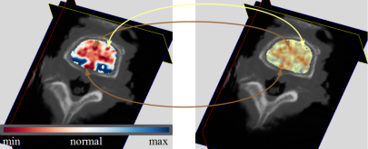
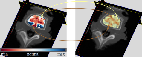
The assessment of bone mineral density (BMD) in vertebrae is critical for the diagnosis of osteoporosis. Recent developments in dual-source CT allow for the simultaneous acquisition of two image data sets with different X-ray tube energies – dual-energy CT (DECT). We have developed a comprehensive approach for assessing the density of the trabecular bone in vertebrae of the spine based on DECT image data.
For this, we have extended an existing biophysical model for computing individual density values for all voxels contained in the trabecular region. Furthermore, we have introduced the barycentric space of the fractional volumes (bone matrix material, adipose and nonadipose tissue) for the purpose of investigating the trabecular bone composition. Based on normal values obtained from the literature, we define a color mapping for the visualization of computed values in 2D and 3D.
S. Wesarg, M. Erdt, K. Kafchitsas, M.F. Khan
CAD of Osteoporosis in Vertebrae Using Dual-energy CT
In: Dillon, Tharam et al. ; 23rd IEEE Symposium on Computer-Based Medical Systems. 2010. IEEE Computer Society, 2010.
S. Wesarg, M. Erdt, K. Kafchitsas, M.F. Khan
Direct visualization of regions with lowered bone mineral density in dual-energy CT images of vertebraeto appear
In: Proc. of SPIE Medical Imaging 2011. 2011.


